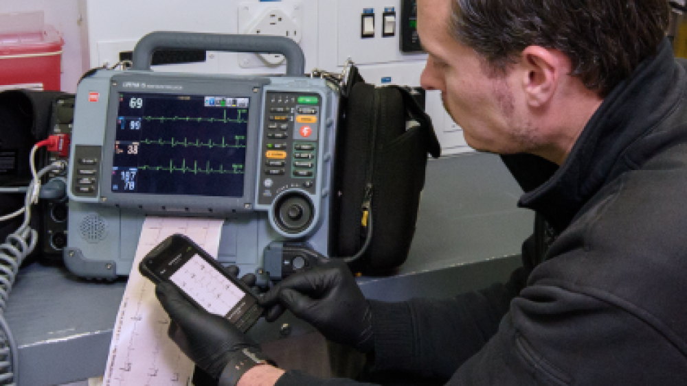
12-lead electrocardiograms (ECGs) have become a mainstay of advanced life support EMS and patient care guidelines call for their acquisition in a wide range of patient complaints. The non-invasive procedure is quick to complete and is relatively inexpensive per procedure once the initial hardware investment has been made. It is common practice now to see 12-lead ECGs acquired for chest pain, difficulty breathing, syncope, general weakness, arrhythmias, abdominal pain and many other complaints.
Missing the mark
Unfortunately, along with the common use of 12-lead ECGs, comes the undesirable side-effect of sloppy electrode placement. Simply guessing or placing the stickers without identifying landmarks to speed up the process gets more common and slowly becomes the norm. Recent observational studies have shown that only 5.8 to 41.6% of paramedics demonstrated correct placement of all the electrodes on manikins [1].
Unfortunately, it is not just EMTs and paramedics losing sight of the proper locations for those 10 little tabs. Hadjiantoni reported in a 2021 article that nurses misplace the leads 64% of the time and cardiologists place leads V1 and V2 in the correct locations in less than 20% of cases [2].
Does lead placement really matter?
Considering how many different shapes and sizes our patients come in, does sliding the leads off the mark one direction or another really change the ECG? It absolutely does. The most well-known variation that can occur is that misplaced V1 and V2 leads may reflect false-positive anterior ST elevation or the appearance of incomplete right bundle branch blocks.
Researchers have also observed that the limb leads are often intentionally placed on the patient’s chest and abdomen instead of on the arms and legs as intended. This may be because it is easier than trying to access the limbs in a fully dressed pre-hospital patient. Or it may occur because EMS cardiac monitors use the same limb leads for continuous monitoring throughout the transport and providers see less artifact when the electrodes are placed more centrally. The harm is that this placement can hide inferior infarcts while creating false-positive lateral infarcts.
Last but not least, consider the issues that may arise if an initial ECG is recorded by EMS using inaccurate lead placement, and then another ECG is obtained in the emergency department with correct electrode positioning. Differences between the two tracings may be mistaken for physiological changes in the patient’s condition, rather than inconsistent techniques.
| More: Become an EKG detective. Focus on what you can identify within an ECG tracing, rather than the criteria
Innovative solution
The methods and technology used for 12 lead ECGs have not changed much in the last 50 years. The electrodes have been improved to better adhere to the patient, but until now, there have been few advances to facilitate proper placement. Two innovative emergency medicine physicians joined forces as CB Innovations, LLC, and received FDA registration in October 2023 for a product that potentially fills this gap. The EXG Radiolucent Electrode System is an ECG electrode sticker that incorporates all 10 of the electrode sites into one adhesive device. The central portion of the EXG is placed over the sternum and a marker on the sticker is aligned at, or slightly below, the nipple line. The right arm, left arm and left leg stickers are attached to the corresponding limbs. Since it only functions as a ground, the right leg lead is incorporated into the central part of the sticker. One more section of the system is stretched under the left breast area and the leads are aligned with mid-clavicular and mid-axillary lines.
The device attaches to a reusable universal adapter to interface with the service’s ECG or multi-function monitor. The EXG can be used during CPR and is radiolucent to allow chest X-rays, CT scans and cardiac cath lab procedures.
The EXG 3L Extension is a 3-lead sticker with an adapter that can be added to the system to acquire right-sided or posterior ECGs.
The EXG 12-lead stickers retail for $10-12 each and the 3L Extension is $5-6. Users will also require one-time purchases of the universal adapter for $100 and a cable for $100. A second cable is required to acquire right side and posterior ECGs.
The EXG is currently available for sale only through CB Innovations, but they will likely be sold through larger distributors in the future. Check them out at future EMS conferences such as EMS World Expo, JEMS@FDIC, and NAEMSP.
Stay safe out there.
REFERENCES
1. Abobaker A, Rana RM. V1 and V2 pericordial leads misplacement and its negative impact on ECG interpretation and clinical care. Annals of Noninvasive Electrocardiology. 2021;26(4). https://www.ncbi.nlm.nih.gov/pmc/articles/PMC8293594/
2. Hadjiantoni A, Oak K, Mengi S, Konya J, Ungvari T. Is the correct anatomical placement of the electrocardiogram (ECG) electrodes essential to diagnosis in the clinical setting: a systematic review. Cardiology and Cardiovascular Medicine. 2021;5(2):182-200. https://www.fortunejournals.com/articles/is-the-correct-anatomical-placement-of-the-electrocardiogram-ecg-electrodes-essential-to-diagnosis-in-the-clinical-setting-a-syste.html
3. Khan, G. M. (2015). A new electrode placement method for obtaining 12-lead ECGs. Open Heart, 2(1). https://doi.org/10.1136/openhrt-2014-000226


