Editor’s note: The EKG Detective will be a monthly column dedicated to illustrating the benefits of utilizing deductive logic as a method for interpreting ECG tracings. The column will highlight and review all the basic ECG interpretations, before transitioning into a monthly interpretation challenge. See you next month and remember, it is always better to practice as a clinician rather than a technician.
Welcome back to the EKG Detective. This column is dedicated to illustrating the benefits of utilizing deductive logic as a method for interpreting EKG tracings. For this month’s article, we will be looking at premature atrial contractions (PACs).
If you need a refresher on inductive and deductive logic, check out our introductory article.
Fill out the form on this page to download your copy of the EKG Detective Interpretation Checklist.
Throughout this series, we will be using the EKG Detective Interpretation Checklist (see Figure 1). This checklist is intended to prompt providers through five sequential elements associated with basic EKG interpretation while working through the specific criteria for each element:
- Rhythm regularity
- Rhythm rate
- P-wave criteria
- PR interval
- QRS criteria
EKG rhythms will be eliminated as we identify criteria within the EKG tracing until there is only one probable interpretation. We will use this checklist to illustrate how deduction is used to interpret an ECG tracing. More practically, it can be used as an EKG interpretation job aid.
Atrial flutter
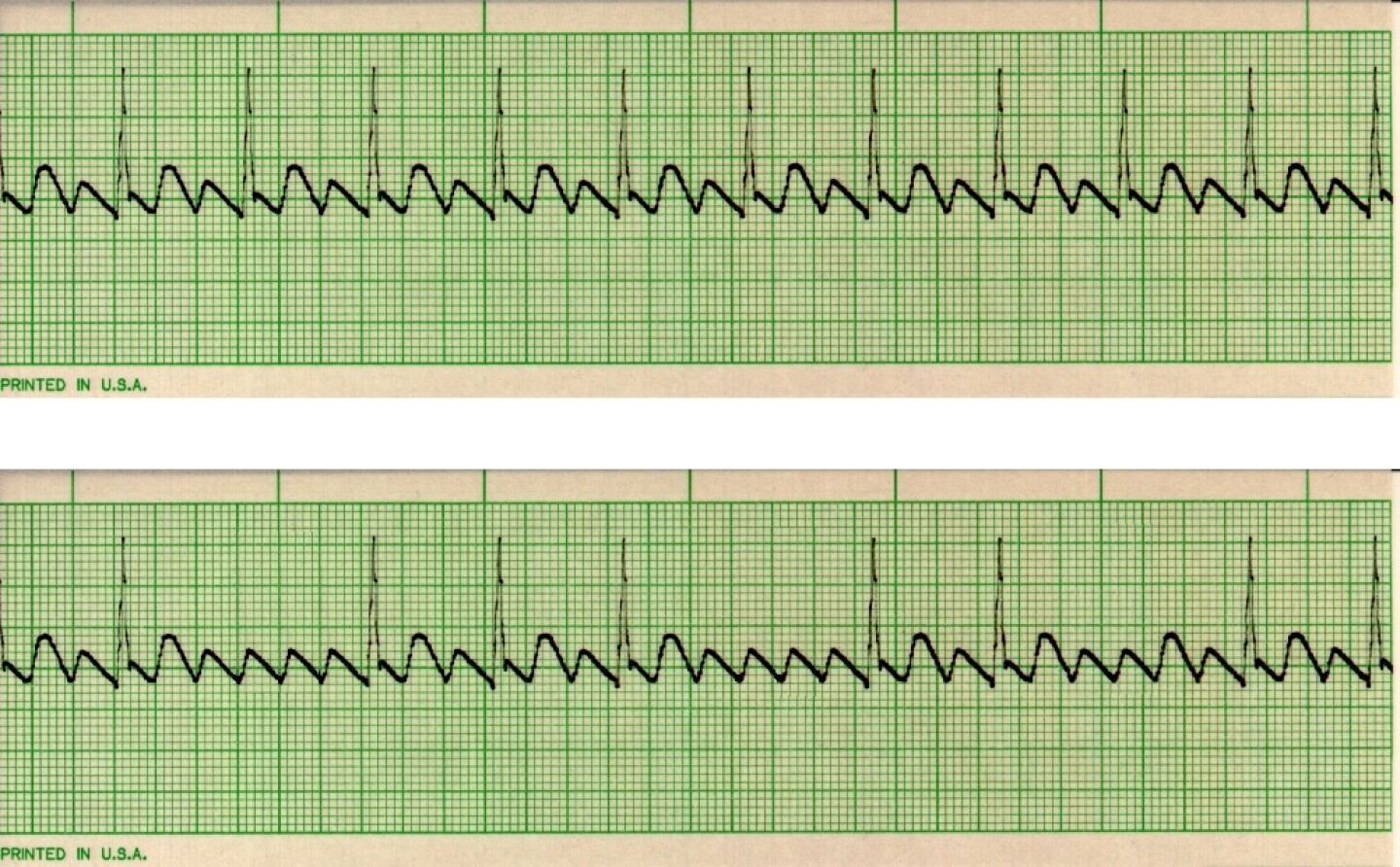
For this article, we will be looking at atrial flutter to illustrate the principle behind the EKG Detective. (See Figure 2 for examples of atrial flutters).
Atrial flutter originates from a single focus within the atria but not within the sinoatrial (SA) node. This is important to note because atrial foci do not have rate limitations that are typically associated with the SA node. Although the SA node can accelerate into the mid to upper 100 beats per minute, it is not designed to sustain those rates for extended periods of time. As a byproduct, the “atrial” rate (P-waves) for atrial flutters beats between 250 to 350 beats per minute and is typically regular and constant.
EKG Category 1: Rhythm regularity
Atrial flutter can present with a regular or irregular ventricular rhythm.
Because the rhythm can be regular or irregular, you will work down the associated side of the EKG Detective Interpretation Checklist that is relevant for the type of rhythm that is present. For illustration’s sake, we will use the atrial flutter that has a regular ventricular rhythm. Since we are working with a regular atrial flutter, we can eliminate any rhythm that is irregular (see Figure 3).
Ectopic beats are uncommon in the setting of atrial flutter. On the outside chance there are ectopic beats, they will present with abnormal P-wave criteria that are typically associated with PACs and PJCs or will have different looking QRS complexes that are associated with PVCs. As there are no ectopic beats within this rhythm, we can eliminate them from the checklist (see Figure 3).
EKG Category 2: Rhythm rate
Atrial flutter cannot be defined by its rate. The ventricular rate typically falls between 60 to 100 beats per minute, but it can also be less than 60 or greater than 100 beats per minute. Due to the variability of ventricular rates for atrial flutter, no rhythms will be eliminated from the checklist based upon this criterion.
EKG Category 3: P-wave criteria
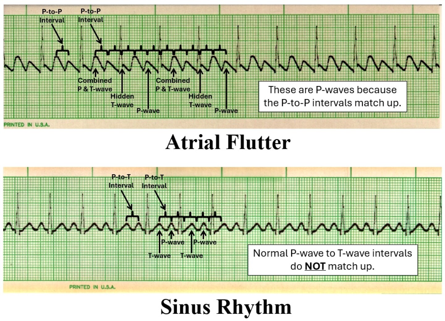
As mentioned, the atrial or P-wave rate for atrial flutter has a regular rhythm and beats between 250 to 350 beats per minute. This is important because the P-wave to P-wave (P-to-P) intervals for an atrial flutter will be similar and match up. Whereas the P-wave to T-wave (P-to-T) intervals within a normal ECG will not be similar or match up.
To determine if the rhythm is atrial flutter, you need to plot out the potential P-to-P interval. This is accomplished by measuring the intervals between what appears to be each separate P-wave, then match that P-to-P interval across the EKG tracing. In atrial flutter, the potential P-to-P intervals will match up. This means P-waves will be combined within T-waves and hidden within QRS complexes (see Figure 4 for examples of matching up intervals).
If the “P-to-P” intervals match up, these P-waves are often referred to as F-waves and/or flutter waves. Many health professionals say atrial flutter waves have a saw-toothed pattern typically seen on the cutting edge of a hand saw. As our EKG example has flutter waves, we will eliminate all the rhythms except for atrial flutter (see Figure 5).
EKG Category 4: PR interval
There is no need to move onto ECG Category 4, PR interval, because you cannot accurately measure a PR interval in the setting of atrial flutter and the only remaining choice on the checklist is the atrial flutter we have been working from.
EKG Category 5: QRS criteria
There is no need to move onto ECG Category 5, QRS Criteria, because the only remaining choice on the checklist is the atrial flutter we have been working from.
Identifying atrial flutter
This example illustrates how deductive logic is used to interpret atrial flutter. The atrial rate typically beats between 250 to 350 beats per minute and is regular. This means the dysrhythmia will reveal extra P-waves in comparison to QRS complexes. Focus on measuring the similar and constant P-to-P intervals. This will reveal the flutter waves or F-waves that are present within the atrial flutter.
See you next month, and remember it is always better to practice as a clinician rather than a technician.


