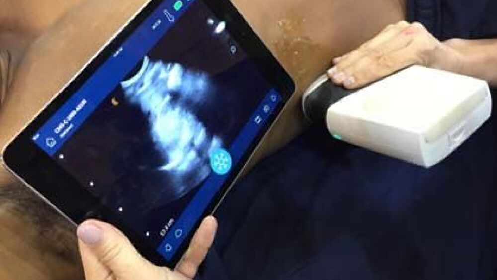
Medic 12 and Engine 14 respond to a report of chest pain. On arrival, the crew finds an elderly male lying in bed looking distressed. The patient’s daughter came over this morning for a visit and found the patient lying on the floor at the bottom of the stairs. She helped him back to the bed and called 911.
The patient complains of right-side chest and abdominal pain. He rates the pain as 4/10 and cannot remember the details of falling. On examination, the patient’s heart rate is 118, his B/P is 98/55 mm Hg, respirations are 34 and non-labored. The room-air pulse oximetry value is 95 percent, oral temperature is 98.2 F and the GCS is 13 (E3, V4, M6). The chest wall is tender on the right side; however, there is marked tenderness and guarding over the right upper quadrant of the abdomen.
Despite the patient’s discomfort, he is adamant that he does not want to go to the hospital. He says he believes he will be fine after some rest. Medic Salazar convinces the patient to let him inspect his abdomen more closely before making any further decisions.
Using a portable ultrasound (which has only been on the ambulance for about a week), Salazar places the probe in the right mid-axillary line at about the level of the ninth intercostal space. After some manipulation of the probe, he recognizes a dark area clearly separating the liver from the right kidney (Morrison’s pouch). With this finding, he is sure the patient is bleeding into the abdomen.
After explaining these findings to the patient and his daughter, the patient finally agrees to go to the hospital. During transport, Salazar establishes an IV and administers a single fluid bolus of 500 mL. The remainder of the transport is uneventful, and the patient arrives safely in the emergency department.
Study review of prehospital sonography
Researchers in Canada performed a systematic review of all published evidence on the question of whether out-of-hospital sonography of the thorax or abdomen changes prehospital diagnosis, treatment or transport destination decisions for the patient [1]. The authors performed a literature search only back to 1990, reasoning that portable ultrasound devices are relatively new to the EMS profession and there would not be much, if any, literature prior to that time. To be included for review, the authors required access to the full paper (as opposed to just an abstract) and that each paper be published in a peer-reviewed journal in the English language.
Although systematic reviews are not primary research reports, they are useful for summarizing available research on a specific issue. When performed well, systematic reviews provide reliable estimates of treatment effects, which may be difficult to estimate from individual studies reviewed in isolation [2].
Results of systematic review
The search identified 992 unique papers, although only eight actually met the inclusion criteria. Of those eight, three had a low risk of bias (error) with a moderate probability of establishing a causal relationship between the use of ultrasound and a change in the prehospital diagnosis, treatment, or destination of the patient [3-5]. The other five studies had a much higher risk of bias, which introduces stronger doubt about a causal relationship between the two variables [6-10].
Six of the studies involved EMS systems located in Europe [4-9], while the remainder involved Australian EMS systems [3,10]. All included both penetrating and blunt trauma in all age groups. Although paramedics were present on the scene in some of the studies, physicians were the primary caregivers in all eight studies.
Collectively, the eight studies suggested that prehospital ultrasound improved the diagnostic accuracy of the caregivers in about two-thirds of all cases. Including ultrasound in the assessment allowed prehospital personnel to rule out injuries (such as hemoperitoneum) or to find injuries missed with traditional exam techniques (such as pneumothorax). In some of those cases, rescuers missed free fluid in the pleural or intraperitoneal cavities during the first ultrasound examination but were able to see the fluid during a repeat examination [4].
Half of the studies reported that the results of the prehospital ultrasound influenced prehospital treatment or destination choices. In some cases, ultrasound imaging assisted in needle aspiration of the pericardial space [10] and suggested the need for chest tube insertion when physical exam techniques failed to identify the injury [8].
In one case, ultrasound supported the ECG interpretation of asystole by confirming the absence of cardiac muscle movement, which resulted in termination of resuscitation efforts in the field [8]. In another case, ultrasound detected the presence of a non-viable fetus, which altered the triage decision for the patient during a mass casualty incident [7].
What these results mean for you
The use of ultrasound is an important component of in-hospital point-of-care (POC) examination for pediatric and adult patients suffering from a traumatic injury with the potential for intra-abdominal or intrathoracic hemorrhage [11,12]. POC ultrasonography can rapidly identify life-threatening injuries that require immediate intervention [13] and has greater sensitivity and specificity for detecting pneumothorax when compared to supine chest radiography [14].
Although patients receiving US in the emergency department (ED) arrive in surgery sooner [15,16] and spend less time in the hospital [16], a recent meta-analysis could find no mortality advantages provided by emergency ultrasound for patients suffering from blunt abdominal trauma [17].
However, the benefits of prehospital ultrasound performed by paramedics for trauma patients are less clear [18]. Much of the prehospital evidence involves either physicians or aeromedical crews performing ultrasound [5,9,10]. In the studies that do involve ground-transport paramedics, the participants often achieve diagnostic accuracy comparable to highly experienced ultrasound experts during training and in simulation [19,20].
In clinical practice, researchers found moderate diagnostic accuracy among nurses and paramedics using ultrasound on a helicopter ambulance in Texas [21]. In this study, when any of the abdominal, cardiac or lung components of the prehospital ultrasound were positive, there was a greater probability of injury than when the prehospital ultrasound examination had negative results. However, sensitivity was not sufficient to rule out injury. Thus, prehospital ultrasound in this case was useful when injury really did exist but had limited value when the ultrasound results were negative.
In contrast, ground-transport paramedics using hand-held ultrasound devices in Minnesota correctly identified every case of intraperitoneal or pericardial fluid when it existed (true positive). In addition, the medics correctly identified the absence of the intraperitoneal or pericardial fluid and the absence of abdominal aneurysm when the condition did not exist (true negative) [22]. This is similar to the results obtained in Germany that demonstrated no difference in diagnostic accuracy between paramedics and emergency department physicians newly trained in the use of ultrasound and those who had routinely used ultrasound in their clinical practice for more than three years [23]. Intermediate-level emergency medical technicians demonstrated diagnostic performance comparable to surgeons and ED physicians when using ultrasound in an urban, tertiary ED to detect intraperitoneal free fluid [24].
Prehospital sonography may also be useful in the assessment and treatment of pneumothorax. One in five patients who suffers a severe traumatic injury has pneumothorax [25]. This condition can rapidly deteriorate into tension pneumothorax. Analysis of trauma registry data from southern Australia suggests that paramedics using traditional assessment techniques may fail to recognize and treat tension pneumothorax in about 37 percent of cases [26]. In a cadaveric model of pneumothorax, out-of-hospital critical care providers using ultrasound correctly identified either the presence (100 percent sensitivity) or absence (100 percent specificity) of sliding lung sign, a marker for pneumothorax [27].
Performing ultrasound examinations in the field is not without its limitations. False negatives for intraabdominal or intrathoracic bleeding can result from ultrasonography being performed before accumulation of blood volumes sufficient for detection [28]. Sonography is less sensitive for low-grade solid organ injuries than for significant intraabdominal free fluid or major hemoperiotenium [29].
Recently in the Paramedic Ultrasound in Cardiac Arrest (PUCA) study, researchers evaluated whether paramedics could obtain satisfactory ultrasonography images of the heart during the 10-second chest compression pause used to deliver ventilation and perform a rhythm and pulse check for patients in cardiac arrest [30]. These images would allow paramedics to determine whether there was any movement of heart muscle or whether the patient had cardiac standstill. Medics were able to obtain an excellent or adequate view on the first attempt in 80 percent of the cases. However, use of ultrasonography produced a median hand-off time of 17 seconds, far longer than the 10 seconds recommended by the American Heart Association [31]. In addition, despite directions to use ultrasonography during every patient encounter involving cardiac arrest, paramedics preferentially used the procedure in case where termination of resuscitation attempts in the field appeared to be the goal.
Limitations of the present study
There are two major limitations in this systematic review that threaten the generalizability of the results. First, the low quality of most of the evidence. The authors rated five of the eight studies used in the analysis as having low-quality evidence with a significant risk of bias. The other three studies had acceptable level of bias, but still did not have evidence quality sufficient to draw definitive conclusions about whether prehospital ultrasound improves patient outcomes.
The second major limitation is that all of the studies occurred either in Europe or in Australia. Physicians staffed all of the ambulances used in each of the reviews, although paramedics accompanied the physicians in at least one of the eight studies. It is difficult to extrapolate success from a physician-staffed ambulance to an EMS system staffed by non-physicians.
Summary
About 4 percent of EMS agencies in the United States currently use ultrasound, although about 21 percent are considering adding the technology [32]. To date, there is limited high-quality evidence that prehospital use of ultrasound improves clinical outcomes for patients suffering a traumatic injury [33]. In spite of this, the overall trend in the available evidence suggests that ultrasound may positively impact individual components of the prehospital patient encounter, including diagnosis, triage and treatment decisions [33].
--
Editor’s Note: The author has no financial interest, arrangement, or direct affiliation with any corporation that has a direct interest in the subject matter of this presentation, including manufacturer(s) of any products or provider(s) of services mentioned.
References
1. O’Dochartaigh, D., & Douma, M. (2015). Prehospital ultrasound of the abdomen and thorax changes trauma patient management: A systematic review. Injury, 46(11), 2093-2102. doi:10.1016/j.injury.2015.07.007
2. Gopalakrishnan, S., & Ganeshkumar, P. (2013). Systematic reviews and meta-analysis: Understanding the best evidence in primary healthcare. Journal of Family Medicine and Primary Care, 2(1), 9–14. doi:10.4103/2249-4863.109934
3. Bodnar, D., Rashford, S., Hurn, C., Quinn, J., Parker, L., Isoardi, K., & Williams, S. (2014). Characteristics and outcomes of patients administered blood in the prehospital environment by a road based trauma response team. Emergency Medicine Journal, 31(7), 583-588. doi:10.1136/emermed-2013-202395
4. Brun, P. M., Bessereau, J., Levy, D., Billeres, X., Fournier, N., & Kerbaul, F. Stay and play eFAST or scoop and run eFAST? That is the question! American Journal of Emergency Medicine, 32(7), 817.e1-817.e2. doi:10.1016/j.ajem.2013.12.063
5. Walcher, F., Weinlich, M., Conrad, G., Schweigkofler, U., Breitkreutz, R., Kirschning, T., & Marzi, I. (2006). Prehospital ultrasound imaging improves management of abdominal trauma. British Journal of Surgery, 93(2), 238–242. doi:10.1002/bjs.5213
6. Busch, M. (2006). Portable ultrasound in pre-hospital emergencies: A feasibility study. Acta Anaesthesiologica Scandinavica, 50(6), 754–758. doi:10.1111/j.1399-6576.2006.01030.x
7. Hoyer, H. X., Vogl, S., Schiemann, U., Haug, A., Stolpe, E., & Michalski, T. (2010). Prehospital ultrasound in emergency medicine: Incidence, feasibility, indications and diagnoses. European Journal of Emergency Medicine, 17(5), 254–259. doi:10.1097/MEJ.0b013e328336ae9e
8. Ketelaars, R., Hoogerwerf, N., & Scheffer, G. J. (2013). Prehospital chest ultrasound by a Dutch helicopter emergency medical service. Journal of Emergency Medicine, 44(4), 811–817. doi:10.1016/j.jemermed.2012.07.085
9. Lapostolle, F., Petrovic, T., Lenoir, G., Catineau, J., Galinski, M., Metzger, J., Chanzy, E., & Adnet, F. (2006). Usefulness of hand-held ultrasound devices in out-of hospital diagnosis performed by emergency physicians. American Journal of Emergency Medicine, 24(2), 237–242. doi:10.1016/j.ajem.2005.07.010
10. Mazur, S. M., Pearce, A., Alfred, S., & Sharley, P. (2007). Use of point-of care ultrasound by a critical care retrieval team. Emergency Medicine Australasia, 19(6), 547–552. doi:10.1111/j.1742-6723.2007.01029.x
11. Diercks, D. B., Mehrotra, A., Nazarian, D. J., Promes, S. B., Decker, W. W., & Fesmire, F. M. (2011). Clinical policy: Critical issues in the evaluation of adult patients presenting to the emergency department with acute blunt abdominal trauma. Annals of Emergency Medicine, 57(4), 387-404. doi:10.1016/j.annemergmed.2011.01.013
12. Marin, J. R., Abo, A. M., Doniger, S. J., Fischer, J. W., Kessler, D. O., Levy, J. A., Noble, V. E., Sivitz, A. B., Tsung, J. M., Vieira, R. L., & Lewiss, R. E. (2015). Point-of-care ultrasonography by pediatric emergency physicians. Policy statement. Annals of Emergency Medicine, 65(4), 472-478. doi:10.1016/j.annemergmed.2015.01.028
13. Whitson, M. R., & Mayo, P. H. (2016). Ultrasonography in the emergency department. Critical Care, 20(1), 227. doi:10.1186/s13054-016-1399-x
14. Gentry Wilkerson, R., & Stone, M. B. (2010). Sensitivity of bedside ultrasound and supine anteroposterior chest radiographs for the identification of pneumothorax after blunt trauma. Academic Emergency Medicine, 17(1), 11–17. doi:10.1111/j.1553-2712.2009.00628.x
15. Ferrada, P., Vanguri, P., Anand, R. J., Whelan, J., Duane, T., Wolfe, L., & Ivatury, R. (2012). Flat inferior vena cava: Indicator of poor prognosis in trauma and acute care surgery patients. American Surgeon, 78(12), 1396-1398.
16. Melniker, L. A., Leibner, E., McKenney, M. G., Lopez, P., Briggs, W. M., & Mancuso, C. A. (2006). Randomized controlled clinical trial of point-of-care, limited ultrasonography for trauma in the emergency department: The first sonography outcomes assessment program trial. Annals of Emergency Medicine, 48(3), 227–235. doi:10.1016/j.annemergmed.2006.01.008
17. Stengel, D., Rademacher, G., Ekkernkamp, A., Güthoff, C., & Mutze, S. (2015). Emergency ultrasound-based algorithms for diagnosing blunt abdominal trauma. Cochrane Database of Systematic Reviews, CD004446. doi:10.1002/14651858.CD004446.pub4
18. McCallum, J., Vu, E., Sweet, D., & Kanji, H. D. (2015). Assessment of paramedic ultrasound curricula: A systematic review. Air Medical Journal, 34(6), 360-368. doi:10.1016/j.amj.2015.07.002
19. Brooke, M., Walton, J., Scutt, D., Connolly, J., & Jarman, B. (2012). Acquisition and interpretation of focused diagnostic ultrasound images by ultrasound-naïve advanced paramedics: Trialling a PHUS education programme. Emergency Medicine Journal, 29(4), 322-326. doi:10.1136/emj.2010.106484
20. Rooney, K. P., Lahham, S., Lahham, S., Anderson, C. L., Bledsoe, B., Sloane, B., Joseph, L., Osborn, M. B., & Fox, J. C. (2015). Pre-hospital assessment with ultrasound in emergencies: Implementation in the field. World Journal of Emergency Medicine, 7(2), 117-123. doi:10.5847/wjem.j.1920-8642.2016.02.006
21. Press, G. M., Miller, S. K., Hassan, I. A., Alade, K. H., Camp, E., Junco, D. D., & Holcomb, J. B. (2014). Prospective evaluation of prehospital trauma ultrasound during aeromedical transport. Journal of Emergency Medicine, 47(6), 638–645. doi:10.1016/j.jemermed.2014.07.056
22. Heegaard, W., Hildebrandt, D., Spear, D., Chason, K., Nelson, B., & Ho, J. (2010). Prehospital ultrasound by paramedics: Results of field trial. Academic Emergency Medicine, 17(6), 624-630. doi:10.1111/j.1553-2712.2010.00755.x
23.Walcher, F., Kirschning, T., Müller, M. P., Byhahn, C., Stier, M., Rüsseler, M., Brenner, F., Braun, J., Marzi, I., & Breitkreutz, R. (2010). Accuracy of prehospital focused abdominal sonography for trauma after a 1-day hands-on training course. Emergency Medicine Journal, 27(5), 345-349. doi:10.1136/emj.2008.059626
24. Kim, C. H., Shin, S. D., Song, K. J., & Park, C. B. (2012). Diagnostic accuracy of Focused Assessment with Sonography for Trauma (FAST) examinations performed by emergency medical technicians. Prehospital Emergency Care, 16(3), 400-406. doi:10.3109/10903127.2012.664242
25. Di Bartolomeo, S., Sanson, G., Nardi, G., Scian, F., Michelutto, V., & Lattuada, L. (2001). A population-based study on pneumothorax in severely traumatized patients. Journal of Trauma, 51(4), 677-682. doi:10.1097/00005373-200110000-00009
26. Heng, K., Bystrzycki, A., Fitzgerald, M., Gocentas, R., Bernard, S., Niggemeyer, L., Cooper, D. J., & Kossmann, T. (2004). Complications of intercostal catheter insertion using EMST techniques for chest trauma. ANZ Journal of Surgery, 74(6), 420-423. doi:10.1111/j.1445-1433.2004.03023.x
27. Lyon, M., Walton, P., Bloch, A., & Shiver, S. A. (2009). 321: Out-of-hospital critical care providers’ retention of ultrasound skills for diagnosis of pneumothoraces: A nine-month followup [abstract]. Annals of Emergency Medicine, 54(3), S100-S101. doi:10.1016/j.annemergmed.2009.06.352
28. Kirkpatrick, A. W., Breeck, K., Wong, J., Hamilton, D. R., McBeth, P. B., Sawadsky, B., & Betzner, M. J. (2005). The potential of handheld trauma sonography in the air medical transport of the trauma victim. Air Medical Journal, 24(1), 34-39. doi:10.1016/j.amj.2004.10.012
29. Kirkpatrick, A. W., Sirois, M., Laupland, K. B., Goldstein, L., Brown, D. R., Simons, R. K., Dulchavsky, S., & Boulanger, B. R. (2005). Prospective evaluation of hand-held focused abdominal sonography for trauma (FAST) in blunt abdominal trauma. Canadian Journal of Surgery, 48(6), 453-460.
30. Reed, M. J., Gibson, L., Dewar, A., Short, S., Black, P., & Clegg, G. R. (2017). Introduction of paramedic led Echo in Life Support into the pre-hospital environment: The PUCA study. Resuscitation, 112, 65-69. doi:10.1016/j.resuscitation.2016.09.003
31. Kleinman, M. E., Brennan, E. E., Goldberger, Z. D., Swor, R. A., Terry, M., Bobrow, B. J., Gazmuri, R. J., Travers, A. H., & Rea, T. (2015). Part 5: Adult basic life support and cardiopulmonary resuscitation quality: 2015 American Heart Association guidelines update for cardiopulmonary resuscitation and emergency cardiovascular care. Circulation, 132(18 Suppl 2), S414-S4135. doi:10.1161/CIR.0000000000000259
32. Taylor, J., McLaughlin, K., McRae, A., Lang, E., & Anton, A. (2014). Use of prehospital ultrasound in North America: A survey of emergency medical services medical directors. BMC Emergency Medicine, 14, 6. doi:10.1186/1471-227X-14-6
33. Canadian Agency for Drugs and Technologies in Health. (2015). Portable ultrasound devices in the pre-hospital setting: A review of clinical and cost-effectiveness and guidelines. Retrieved from https://www.ncbi.nlm.nih.gov/pubmedhealth/PMH0085902/


