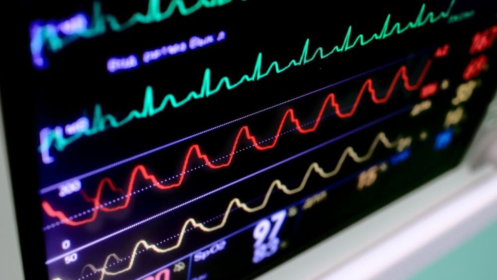
We’ve all heard the saying “treat the patient, not the monitor.” Well, that might be true in most instances, but there are certainly times where the patient looks fine, but really isn’t.
You read that correctly; treat the monitor, not just the patient.
It seems counterintuitive to what has been preached in every paramedic class (and even in some EMT classes, too).
In this context, we often refer to a sick appearing patient who has a seemingly normal monitor display; ECG rhythm is normal, blood pressure is normal, pulse oximetry, etc. But, what about the instances where the coin is flipped – where the patient appears normal, but the monitor is screaming at you: sick!?
While there are certainly instances where the monitor may sway your thought process down some maze of what ifs – especially when your patient appears seemingly stable – there are a handful of times where we need to react based upon what our monitor tells us. In some situations, it may be the precursor to a rapidly deteriorating patient (they just don’t know it yet!).
Stable STEMI
Your patient presents with chest pain or discomfort, jaw pain, upper back pain or a shooting pain down his arm ... sounds like an acute coronary syndrome, right?
Limb leads go on the limbs (every time!), precordial leads on the chest. “Sir, can you sit still for about 15 seconds for me, please?”
Your patient appears to have normal skin color, condition and temperature. His blood pressure is normotensive, heart rate is in the 70s with an underlying normal sinus rhythm, and his pulse oximetry is well above 95% with no signs of dyspnea noted. Your patient appears stable.
His diagnostic 12-lead ECG print-out, however, tells you otherwise. If he had a “classic” symptom of a heart attack, this would all come together perfectly. But, let’s say he didn’t. Let’s say his only symptom was weakness, or even acute malaise. Now what? His physical presentation tells you that he’s stable, but his ECG tells you he’s sick (emergently sick).
Not only is this a prime example of why 12-lead ECG interpretation is absolutely warranted in any patient with a pulse and a problem, it’s a prime example of why we need to factor-in what the monitor tells us. If we hadn’t, this patient might have otherwise gone unnoticed, or could have been brushed off as having flu-like symptoms.
Walking with V-Tach
I can recall responding to a residence of a 70s male for weakness symptoms. It was around 23:00 and the patient was found in bed. He was alert, oriented, and his skin was pink, warm and dry. He complained of acute weakness and the inability to fall asleep.
Could this have been brushed off as any number of acute, urgent (not emergent) conditions? Sure.
He appeared completely stable; his symptoms didn’t stand out as alarming. Palpating his pulse, it felt as though it was around 100 and irregular. His manual blood pressure was difficult to obtain (faint sounds), but his palpated blood pressure was around 110/P.
I decided to check out his 12-lead ECG (as relying solely on a rhythm strip can be a costly mistake!). To my disbelief, a wide-complex tachycardia appeared with a rate of approximately 200 complexes per minute. The rhythm was regular in appearance and had concordant morphologies in leads V1 through V6. This was V-Tach (ventricular tachycardia) and my patient was still talking to me (this was my first encounter of an alive V-Tach patient).
Again, based on the patient’s presentation and current symptoms, many EMS providers might have brushed this off as something seemingly benign. By all means, the patient was stable in his condition, but he still warranted ALS treatment (just not immediately going down the route to electricity).
Acute tachycardia
Your patient is intubated following an RSI/DSI procedure and is being mechanically ventilated by your portable ventilator device. As they’re chemically sedated and paralyzed, obtaining any direct information from the patient going forward is, essentially, not possible. As a result, you have to rely on your monitor for different cues related to their current status.
Aside from their paralytic medication wearing off, your patient won’t be able to move (or even breathe) on their own. As such, you need to monitor the patient’s pulse oximetry levels for signs of hypoxia, capnograph for signs of extubation or airway compromise, and heart rate for signs of distress.
If your patient remains chemically paralyzed, but her sedation medication is wearing off, how would you know? The patient certainly can’t tell you this, and simply administering more rocuronium isn’t the right answer.
Look to the heart rate; tachycardia in particular. A sudden change in any vital sign should indicate some form of distress. The patient’s elevated heart rate, as an example, may indicate that their sedative medication is wearing off, and they’re now experiencing a period of panic because they don’t know what’s happening, there’s a tube in their mouth, and they know that they should be breathing on their own, but they can’t. In this case, the patient couldn’t tell us anything, but the monitor could!
Hypocapnea
A recent fever, heart rate of 100, congested cough with respiratory rate around 20, and a seemingly normotensive blood pressure could drive your differential diagnosis down a few different avenues.
It could be COVID-19, pneumonia exacerbation, the flu, a generalize illness or even sepsis. Is CPAP, rest, fluids or antibiotics indicated?
How about assessing the monitor? Sinus tachycardia with a rate of 100, pulse oximetry at 92% on room air, end-tidal carbon dioxide at 23 mmHg with a normal appearing waveform. Herein lies the answer: hypocapnia!
Sure, this could be related to the acutely high respiratory rate of 20, but to that same effect, it would seem more reasonable for the end-tidal measurement to be about 30 mmHg, not 23. That’s more of a hypoperfusion problem. This sounds a lot more like sepsis, above all others.
Treating the monitor, not just the patient, can help to guide your clinical decision-making process toward a more accurate and definitive differential diagnosis. Of course, you still need to look at your patient, touch your patient, and talk to your patient, but your patient doesn’t always have all of the answers. Sometimes, you need to utilize your tools to assist you along the way – like your cardiac and other diagnostic monitors.
Regardless of your scope of practice, utilizing your monitors to help guide your decisions toward an appropriate treatment plan is all part of the big clinical picture. As a clinician, sometimes you need to treat the monitor, not just the patient.
Read next: 5 steps to an accurate physical exam


