Accurate blood pressure measurement is critical for EMS providers making treatment decisions in the field. However, simple mistakes — like using the wrong cuff size or positioning the patient incorrectly — can lead to false readings, potentially impacting patient care. This article outlines 5 common errors that can cause inaccurate blood pressure readings and offers practical tips to improve BP measurement accuracy in prehospital settings.
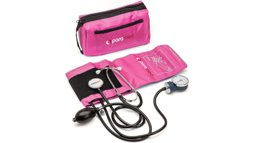
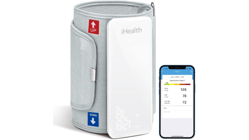
Blood pressure is measured using a sphygmomanometer, which consists of an inflatable cuff, a pressure gauge, and a stethoscope or electronic sensor. There are two main types: manual blood pressure monitors and digital blood pressure monitors. Manual devices, including aneroid sphygmomanometers, require a trained provider to use a stethoscope to listen for blood flow sounds while inflating and deflating the cuff. Digital blood pressure monitors, commonly used at home and in medical settings, automatically inflate the cuff and provide a digital reading of systolic and diastolic pressure. Some advanced models also include features like irregular heartbeat detection and wireless connectivity for tracking blood pressure trends over time.
To make the best use of blood pressure monitoring equipment, it is helpful to have an insight into how the equipment works and the likely sources of error that can affect readings. Download your copy by completing the “Get Access to this EMS1 Resource” box on this page!
What is normal blood pressure?
The American Heart Association published the following healthy and unhealthy blood pressure ranges.
Obtaining accurate blood pressure readings
Blood pressure is measured using two key numbers:
- Systolic blood pressure (top number) measures the force of blood against artery walls when the heart beats.
- Diastolic blood pressure (bottom number) measures the pressure in the arteries when the heart rests between beats.
Both systolic and diastolic blood pressure readings are important in diagnosing high blood pressure (hypertension). However, systolic blood pressure is a stronger predictor of heart disease risk, especially for adults over 50. As people age, systolic blood pressure tends to increase due to:
- Stiffening arteries
- Plaque buildup (atherosclerosis)
- Higher rates of cardiovascular disease
Monitoring blood pressure levels is crucial for maintaining heart health and preventing hypertension-related complications.
During cuff deflation, you will hear Korotkoff sounds through your stethoscope.
They occur in 5 phases:
- First detectable sounds, corresponding to appearance of a palpable pulse
- Sounds become softer, longer and may occasionally transiently disappear
- Change in sounds to a thumping quality (loudest)
- Pitch intensity changes and sounds become muffled
- Sounds disappear
The AHA recommends that clinicians record the systolic BP at the start of phase 1 and the diastolic BP at start of phase 5 Korotkoff sounds [2].
What factors can cause an incorrect blood pressure reading?
The most common blood pressure reading mistakes are:
- Using the wrong-sized cuff
- Incorrect patient positioning
- Incorrect cuff placement
- Normal reading prejudice
- Not factoring in electronic units correctly
What causes false high blood pressure readings?
BP readings can be falsely high due to several factors, including:
- Incorrect cuff size. Using a cuff that is too small can artificially elevate the reading.
- Improper cuff placement. Placing the cuff too low on the arm or not wrapping it snugly can cause inaccurate readings.
- Patient positioning. If the limb is below heart level or unsupported, BP may appear higher than it actually is.
- Fear or anxiety. Anxiety or stress, especially in a clinical setting, can temporarily raise BP.
- Talking or moving. The patient should be still and silent during measurement.
- Recent physical activity. Exercise or exertion immediately before the reading can cause temporary elevation.
- Full bladder. A full bladder can increase systolic BP by 10-15 mmHg.
- Smoking, caffeine, or alcohol. Consuming these within 30 minutes before measurement can artificially raise BP.
- Incorrect inflation or deflation rate. Deflating the cuff too quickly or too slowly can lead to false readings.
- Crossed legs. This can increase systolic BP by 2-8 mmHg.
What causes false low blood pressure readings?
A BP reading can be falsely low due to several factors, including:
- Incorrect cuff size. Using a cuff that is too large can underestimate BP.
- Improper cuff placement. Placing the cuff too high on the limb or loosely wrapping it can result in a lower reading.
- Limb position above heart level. If the arm is too high, gravity can falsely lower BP.
- Failure to support the arm. A relaxed, unsupported arm may cause a lower reading.
- Rapid cuff deflation. Deflating too quickly may lead to missing the true systolic pressure.
- Venous pooling. If the patient has been lying down or sitting too long before the reading, blood may pool in the extremities, leading to lower BP.
- Dehydration or hypovolemia. Low circulating blood volume can cause an inaccurately low BP.
- Cold environment. Peripheral vasoconstriction in response to cold can lead to lower BP readings.
- Background noise. If the provider has difficulty hearing Korotkoff sounds, they may record a falsely low reading.
- Slow inflation of the cuff. This can lead to venous congestion and an inaccurate diastolic reading.
Here’s what many of us do wrong, and how to take a blood pressure reading:
1. You’re using the wrong-sized cuff
The most common error providers make when measuring blood pressure using indirect equipment is using an incorrectly sized cuff. A BP cuff that is too large will give falsely low readings, while an overly small cuff will provide readings that are falsely high.
The American Heart Association publishes guidelines for blood pressure measurement recommending that the bladder length and width (the inflatable portion of the cuff) should be 80% and 40% respectively, of arm circumference [2]. Most practitioners find measuring bladder and arm circumference to be overly time-consuming, so they don’t do it.
The most practical way to quickly and properly size a BP cuff is to pick a cuff that covers two-thirds of the distance between your patient’s elbow and shoulder. Carrying at least three cuff sizes (large adult, regular adult and pediatric blood pressure cuffs) will fit the majority of the adult population. Multiple smaller sizes are needed if you frequently treat pediatric patients.
2. You’ve incorrectly positioned your patient’s body
The second most common error in BP measurement is incorrect limb position. To accurately assess blood flow in an extremity, influences of gravity must be eliminated.
The standard reference level for measurement of blood pressure by any technique — direct or indirect — is at the level of the heart. When using a cuff, the arm (or leg) where the cuff is applied must be at mid-heart level. Measuring BP in an extremity positioned above heart level will provide a falsely low BP whereas falsely high readings will be obtained whenever a limb is positioned below heart level. Errors can be significant — typically 2 mmHg for each inch the extremity is above or below heart level. A seated upright position provides the most accurate blood pressure, as long as the arm in which the pressure is taken remains at the patient’s side. Patients lying on their side, or in other positions, can pose problems for accurate pressure measurement.
To correctly assess BP in a side lying patient, hold the BP cuff extremity at mid heart level while taking the pressure.
In seated patients, be certain to leave the arm at the patient’s side. Arterial pressure transducers are subject to similar inaccuracies when the transducer is not positioned at mid-heart level. This location, referred to as the phlebostatic axis, is located at the intersection of the fourth intercostal space and mid-chest level (halfway between the anterior and posterior chest surfaces. Note that the mid-axillary line is often not at mid-chest level in patients with kyphosis or COPD, and therefore should not be used as a landmark. Incorrect leveling is the primary source of error in direct pressure measurement with each inch the transducer is misleveled causing a 1.86 mmHg measurement error. When above the phlebostatic axis, reported values will be lower than actual; when below the phlebostatic axis, reported values will be higher than actual.
3. You’ve placed the cuff incorrectly
The standard for blood pressure cuff placement is the upper arm using a cuff on bare skin with a stethoscope placed at the elbow fold over the brachial artery. The patient should be sitting, with the arm supported at mid heart level, legs uncrossed, and not talking. Measurements can be made at other locations such as the wrist, fingers, feet, and calves but will produce varied readings depending on distance from the heart. The mean pressure, interestingly, varies little between the aorta and peripheral arteries, while the systolic pressure increases and the diastolic decreases in the more distal vessels.
Crossing the legs increases systolic blood pressure by 2 to 8 mm Hg. About 20% of the population has differences of more than 10 mmHg pressure between the right and left arms. In cases where significant differences are observed, treatment decisions should be based on the higher of the two pressures.
4. Your readings exhibit ‘prejudice’
Prejudice for normal readings significantly contributes to inaccuracies in blood pressure measurement. No doubt, you’d be suspicious if a fellow EMT reported blood pressures of 120/80 on three patients in a row. As creatures of habit, human beings expect to hear sounds at certain times and when extraneous interference makes a blood pressure difficult to obtain, there is considerable tendency to “hear” a normal blood pressure.
Orthostatic hypotension is defined as a decrease in systolic blood pressure of 20 mm Hg or more, or diastolic blood pressure decrease of 10 mm Hg or more measured after three minutes of standing quietly.
There are circumstances when BP measurement is simply not possible. For many years, trauma resuscitation guidelines taught that rough estimates of systolic BP (SBP) could be made by assessing pulses. Presence of a radial pulse was thought to correlate with an SBP of at least 80 mm Hg, a femoral pulse with an SBP of at least 70, and a palpable carotid pulse with an SBP over 60. In recent years, vascular surgery and trauma studies have shown this method to be poorly predictive of actual blood pressure [3].
Noise is a factor that can also interfere with BP measurement. Many ALS units carry doppler units that measure blood flow with ultrasound waves. Doppler units amplify sound and are useful in high noise environments.
BP by palpation or obtaining the systolic value by palpating a distal pulse while deflating the blood pressure cuff generally comes within 10 – 20 mmHg of an auscultated reading. A pulse oximeter waveform can also be used to measure return of blood flow while deflating a BP cuff, and is as accurate as pressures obtained by palpation.
In patients with circulatory assist devices that produce non-pulsatile flow such as left ventricular assist devices (LVADs), the only indirect means of measuring flow requires use of a doppler.
The return of flow signals over the brachial artery during deflation of a blood pressure cuff in an LVAD patient signifies the mean arterial pressure (MAP). While a normal MAP in adults ranges from 70 to 105 mmHg, LVADs do not function optimally against higher afterload, so mean pressures of less than 90 are often desirable.
Clothing, patient access, and cuff size are obstacles that frequently interfere with conventional BP measurement. Consider using alternate sites such as placing the BP cuff on your patient’s lower arm above the wrist while auscultating or palpating their radial artery. This is particularly useful in bariatric patients when an appropriately sized cuff is not available for the upper arm. The thigh or lower leg can be used in a similar fashion (in conjunction with a pulse point distal to the cuff).
All of these locations are routinely used to monitor BP in hospital settings and generally provide results only slightly different from traditional measurements in the upper arm.
5. You’re not factoring in electronic units correctly
Electronic blood pressure units also called Non Invasive Blood Pressure (NIBP) machines, sense air pressure changes in the cuff caused by blood flowing through the BP cuff extremity. Sensors estimate the Mean Arterial Pressure (MAP) and the patient’s pulse rate. Software in the machine uses these two values to calculate the systolic and diastolic BP.
To ensure accuracy from electronic units, it is important to verify the displayed pulse with an actual patient pulse. Differences of more than 10% will seriously alter the unit’s calculations and produce incorrect systolic and diastolic values on the display screen.
Given that MAP is the only pressure actually measured by an NIBP, and since MAP varies little throughout the body, it makes sense to use this number for treatment decisions.
A normal adult MAP ranges from 70 to 105 mmHg. As the organ most sensitive to pressure, the kidneys typically require an MAP above 60 to stay alive, and sustain irreversible damage beyond 20 minutes below that in most adults. Because individual requirements vary, most clinicians consider a MAP of 70 as a reasonable lower limit for their adult patients.
Increased use of NIBP devices, coupled with recognition that their displayed systolic and diastolic values are calculated while only the mean is actually measured, have led clinicians to pay much more attention to MAPs than in the past. Many progressive hospitals order sets and prehospital BLS and ALS protocols have begun to treat MAPs rather than systolic blood pressures.
Finally, and especially in the critical care transport environment, providers will encounter patients with significant variations between NIBP (indirect) and arterial line (direct) measured blood pressure values.
Mitigate NIBP and auscultating innacuracies by watching the plethysmography waveform on your pulse oximeter and noting the mean arterial pressure.
References:
- James PA, Oparil S, Carter BL, et al. 2014. Evidence-Based Guideline for the Management of High Blood Pressure in Adults: Report From the Panel Members Appointed to the Eighth Joint National Committee (JNC 8). JAMA. 2014;311(5):507-520. (Available at: http://jama.jamanetwork.com/article.aspx?articleid=1791497)
- Pickering TG, Hall JE, Appel LJ, et al. AHA Scientific Statement: Recommendations for blood pressure measurement in humans and experimental animals, part 1: blood pressure measurement in humans. Hypertension. 2005; 45: 142-161. (Available at: https://hyper.ahajournals.org/content/45/1/142.full)
- Deakin CD, Low JL. Accuracy of the advanced trauma life support guidelines for predicting systolic blood pressure using carotid, femoral, and radial pulses: observational study. BMJ. 2000; 321(7262): 673–674. (Available at: http://www.ncbi.nlm.nih.gov/pmc/articles/PMC27481/)
- Lehman LH, Saeed M, Talmor D, Mark R, Malhotra A. Methods of blood pressure measurement in the ICU. Crit Care Med. 2013;41:34-40.
This article, originally posted Apr. 9, 2014, has been updated to include additional information and a blood pressure range chart.


